Sonography

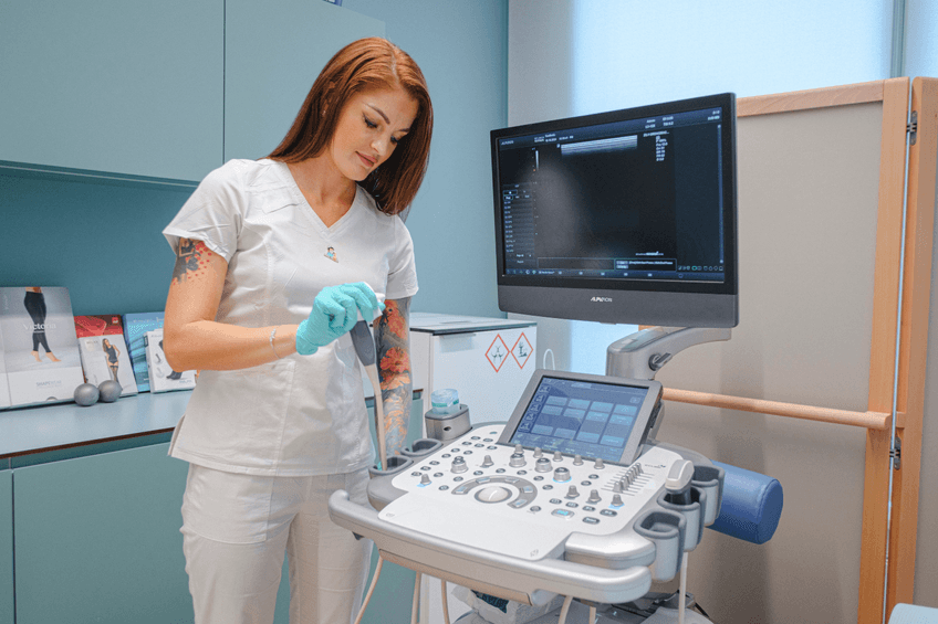
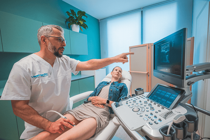
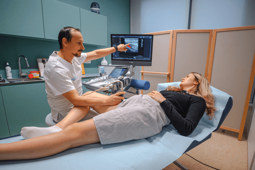
Quality diagnostics is essential in solving any problem. Sonography is an effective imaging method that uses high-frequency ultrasound waves to anatomically image tissues and organs in the area under examination. It is used as a diagnostic tool for both preventive examinations and in the treatment of already diagnosed health problems.
Make an appointmentWho will help you?
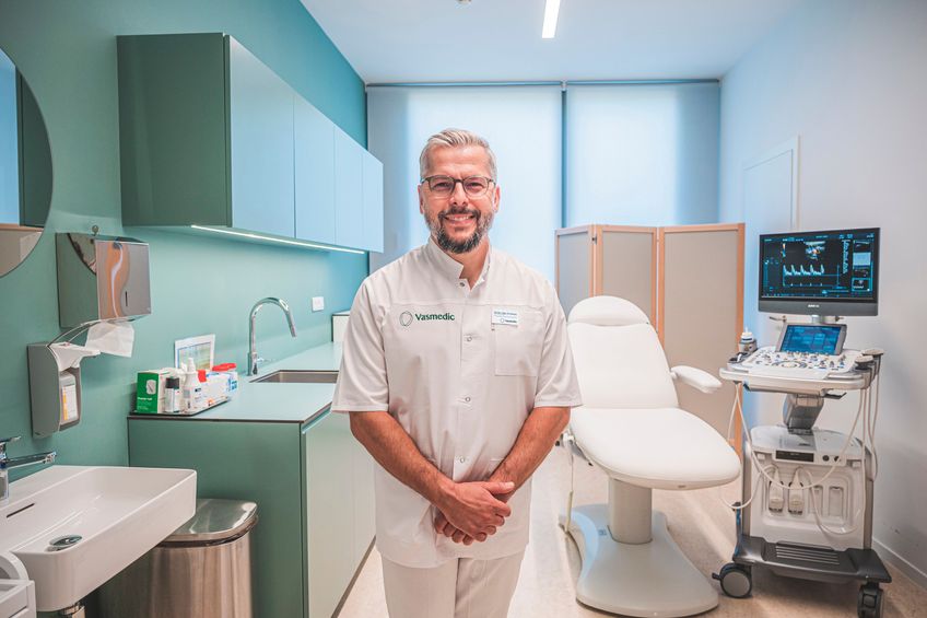
The medical team at Vasmedic consists of leading experts who will provide you with professional care with a personal touch.



See what it looks like here
Practical information for clients
Do you have any questions? Here you will find answers that will make your first step towards care with us easier.
You can book in several ways – via the inquiry form on the website, using the online booking system, by phone or in person at the clinic reception. No referral from a general practitioner or other doctor is needed, you can book directly. Vasmedic Clinic is a private healthcare facility for self-payers.
You can pay for services in cash, by card or by bank transfer. We also offer gift vouchers in several values that can be used for health and aesthetic services. Ideal as a gift for someone close to you.
If you are unable to attend, please let us know as soon as possible – ideally at least 48 hours in advance. This will allow us to offer the appointment to someone else. Late cancellations or no-shows without an excuse may incur a fee.
Pricelist
| Description | Price |
|---|---|
| Ultrasound examination of the abdominal cavity | CZK 2 200 |
| Ultrasound examination of the carotid arteries and branches of the aortic arch | CZK 2 200 |
| Ultrasound examination of the abdominal aorta and its branches | CZK 2 200 |
| Ultrasound examination of the peripheral arteries of the extremities | CZK 2 200 |
| Ultrasound examination of two areas | CZK 3 000 |
| Ultrasound examination of three or more areas | CZK 4 000 |
| Ultrasound examination of another area | CZK 2 200 |
| Control examination | CZK 1 000 |
